


Hip joint anatomy.
Question : Describe the hip joint anatomy?
 Ligaments of the hip joint :-


Answer : The hip joint is a ball and socket synovial joint, formed by an articulation between the pelvic acetabulum and the head of the femur.
It forms a connection from the lower limb to the pelvic girdle, and thus is designed for stability and weight-bearing – rather than a large range of movement.
In this article, we shall look at the anatomy of the hip joint – its articulating surfaces, ligaments and neurovascular supply.
Structure of the hip joint :
Articulating Surfaces
The hip joint consists of an articulation between the head of femur and acetabulum of the pelvis.
The acetabulum is a cup-like depression located on the inferolateral aspect of the pelvis. Its cavity is deepened by the presence of a fibrocartilaginous collar – the acetabular labrum. The head of femur is hemispherical, and fits completely into the concavity of the acetabulum.
Both the acetabulum and head of femur are covered in articular cartilage, which is thicker at the places of weight bearing.
The capsule of the hip joint attaches to the edge of the acetabulum proximally. Distally, it attaches to the intertrochanteric line anteriorly and the femoral neck posteriorly.

Ligaments
The ligaments of the hip joint act to increase stability. They can be divided into two groups – intracapsular and extracapsular:
Intracapsular
The only intracapsular ligament is the ligament of head of femur. It is a relatively small structure, which runs from the acetabular fossa to the fovea of the femur.
It encloses a branch of the obturator artery (artery to head of femur), a minor source of arterial supply to the hip joint.
Extracapsular
There are three main extracapsular ligaments, continuous with the outer surface of the hip joint capsule:


- Iliofemoral ligament – arises from the anterior inferior iliac spine and then bifurcates before inserting into the intertrochanteric line of the femur.
- It has a ‘Y’ shaped appearance, and prevents hyperextension of the hip joint. It is the strongest of the three ligaments.
- Pubofemoral – spans between the superior pubic rami and the intertrochanteric line of the femur, reinforcing the capsule anteriorly and inferiorly.
- It has a triangular shape, and prevents excessive abduction and extension.
- Ischiofemoral– spans between the body of the
- ischium and the greater trochanter of the femur, reinforcing the capsule posteriorly.
- It has a spiral orientation, and prevents hyperextension and holds the femoral head in the acetabulum.
Neurovascular Supply
The arterial supply to the hip joint is largely via the medial and lateral circumflex femoral arteries – branches of the profunda femoris artery (deep femoral artery). They anastomose at the base of the femoral neck to form a ring, from which smaller arteries arise to supply the hip joint itself.The medial circumflex femoral artery is responsible for the majority of the arterial supply (the lateral circumflex femoral artery has to penetrate through the thick iliofemoral ligament). Damage to the medial circumflex femoral artery can result in avascular necrosis of the femoral head.The artery to head of femur and the superior/inferior gluteal arteries provide some additional supply.The hip joint is innervated primarily by the sciatic, femoral and obturator nerves. These same nerves innervate the knee, which explains why pain can be referred to the knee from the hip and vice versa.By TeachMeSeries Ltd (2020)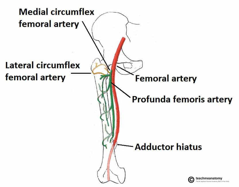 Fig 2 – The medial and lateral circumflex femoral arteries are the major blood supply to the hip joint.
 - ischium and the greater trochanter of the femur, reinforcing the capsule posteriorly.


No comments: